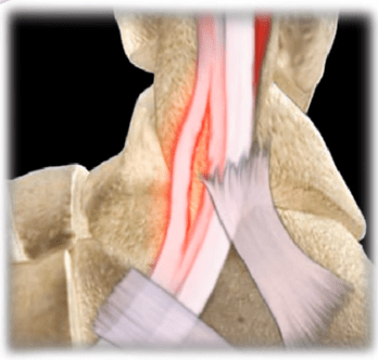
The peroneal tendons run on the outside of the ankle just behind the fibula. The two peroneal tendons lie behind the fibula in the groove. The two peroneal tendons are: the peroneus brevis tenon and the peroneus longus tendon. Patients with peroneal tendon subluxation usually describe pain in the outer part of the ankle or just behind the lateral malleolus. Retromalleolar swelling with pain and giving way of the ankle should alert the clinician to the possibility of peroneal tendon subluxation. The patient may feel a “pop” and click on the outer side of the ankle. Testing for peroneal tendon subluxation is usually done with dorsiflexion/eversion of the foot against resistance. The ankle may feel as if it is unstable and sometimes the patient will be able to demonstrate the subluxation of the tendons. The peroneal tendons are held in their position behind the fibula predominantly by the superior peroneal retinaculum. Peroneal tendon subluxation usually occurs more in younger individuals. It is usually a sports related injury such as with soccer and skiing and the injury can be missed. The condition may be acute or chronic recurrent. It may be associated with a shallow fibular groove or a weak superior peroneal retinaculum. The retinaculum is usually peeled off of the bone due to the injury.
An x-ray may show an avulsion fracture of the fibula, called a “rim fracture”. The Fleck Sign is an indication for peroneal tendon subluxation. An MRI or ultrasound is very helpful in viewing the peroneal tendon subluxation. The condition may be associated with a longitudinal tear of the peroneus brevis tendon (the tendon is near the fibula and may hit the fibula as it subluxes, causing a tear). Treatment for acute subluxation is usually treated with a cast or immobilization. The results are typically average. Surgery to repair the retinaculum is done is there is a rim fracture or in conjuction with other conditions such as a calcaneal fracture. For chronic recurrent and painful dislocations, a soft tissue procedure will be performed to reconstruct the superior peroneal retinaculum. The repair of the longitudinal tear of the peroneus brevis by suturing the tendon side to side.
